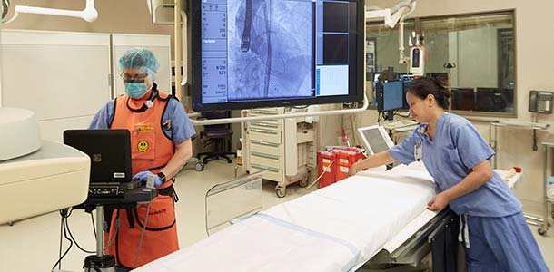
During a cardiac catheterization, interventional cardiologists thread a thin catheter through blood vessels and into the coronary arteries. Using X-rays, they look for signs of heart disease and determine the size and location of fat and calcium deposits that may narrow the openings in the arteries.
The George Washington University Hospital offers some of the most technologically advanced patient care equipment in the country including the Integris Allura, a large field-of-view catheterization lab with three-dimensional imaging. This superior technology provides faster and more precise cardiovascular exams, potentially decreasing time spent in the operating room or saving a patient from surgery altogether.
Integris Allura provides "head-to-toe” imaging of not only the carotid and coronary vessels, but of peripheral vessels as well. As a result, patients no longer need to visit two labs — the cardiac cath lab and the vascular lab — to get a complete imaging picture. The system can be used for all types of heart and vascular diagnostic tests. It is also used for minimally invasive or interventional treatments and surgery, such as coronary stenting, diagnostic catheterization, balloon angioplasty and embolization.
Cardiac Catheterization Treatments
- Transradial Cardiac Catheterization (through the wrist): GW Hospital is one of a few hospitals in the area to offer this alternate approach that has a lower risk of bleed complications and is generally more comfortable for the patient. Learn more about Transradial Cardiac Catheterization>
- Angioplasty: Instead of open-heart surgery, angioplasty uses a a balloon tipped catheter to unblock a coronary artery. Stents, small, flexible tube made of plastic or wire mesh, are sometimes used to support the damaged artery walls. Learn more about Angioplasty
- Atherectomy: Similar to angioplasty, this surgical procedure uses a laser beam or a whirling blade to cut the plaque build-up on the artery wall. This increases blood flow in narrowed arteries and stents also may be implanted during this procedure.
-
Thrombectomy / Thrombolysis: The most effective procedure to treat deep vein thrombosis (DVT), a catheter-directed thrombolysis is performed under imaging guidance by interventional radiologists. DVT is a blood clot commonly found in the lower leg, thigh or deep veins of the pelvis. The clot may travel through the blood stream and potentially lodge in the brain, lungs, heart or other area, causing severe damage.
Transcatheter Aortic Valve Replacement (TAVR)
Transcatheter aortic valve replacement (TAVR) is a minimally invasive procedure for high-risk patients with aortic stenosis who are not candidates for traditional aortic valve surgery. TAVR is a catheter delivered aortic heart valve replacement. Learn more about TAVR
Intracoronary Ultrasound
This procedure uses high frequency sound waves, or ultrasound, to evaluate the heart and determine different treatment needs. A miniature sound probe (transducer) is placed on the tip of a catheter and threaded through the coronary arteries to the heart where it emits sound waves to create the images.
Endomyocardial Biopsy
Used mainly to diagnosis cardiomyopathy, this test involves cutting or scraping a small piece of heart tissue in order to examine the sample closely and detect any abnormalities.
Evaluation of Pulmonary Hypertension
Elevated pressures in the pulmonary arteries can be caused by a variety of conditions. In some patients, shortness of breath can be debilitating. While there are several treatment options, some options are best evaluated by testing response to medication during a right heart catheterization performed in the cardiac cath laboratory. These tests help the pulmonologists optimize patient treatment.
Advanced Devices
The following devices are implanted in the cardiac cath laboratory:
Cardiac-Assist Device
The Abiomed Impella cardiac assist device (the world's smallest ventricle heart pump) provides partial circulatory support for up to six hours in critically ill patients. The device is inserted through the femoral artery in the groin area and with the help of a guide wire is advanced into the left ventricle.
Atrial Septal Defect (ASD) Closure
An atrial septal defect is an abnormal opening in the wall (septum) that divides the two upper chambers of the heart (atria). Individuals with ASD are at an increased risk for developing a number of complications including:
- Atrial fibrillation (in adults)
- Heart failure
- Pulmonary overcirculation
- Pulmonary hypertension
- Stroke
If the defect is small and symptomatic, patients may not need treatment. However for larger ASD openings, when the heart is enlarged or patients are experiencing symptoms, treatment may be recommended. At GW Hospital, physicians implant an atrial septal device to correct this abnormal opening. This is done in the catheterization laboratory and involves placing an ASD closure device into the heart through tubes called catheters to seal off the opening. Patients will typically go home the same day as the procedure.
Designated STEMI Center
We’re Saving Time So We Can Save Lives
During a heart attack, every minute counts. The George Washington University Hospital offers 24-hour interventional cardiology coverage, making it one of only three hospitals in Washington D.C. that has been designated for EMS transport of patients with a type of suspected heart attack known as a STEMI (ST-Segment Elevation Myocardial Infarction).
GW Hospital’s catheterization door-to-balloon times (the time it takes for a cardiac patient to proceed from hospital entrance to catheterization procedure) averages 68 minutes* and 87 percent of our patients are treated in less than 90 minutes*, better than the American College of Cardiology benchmark of 75 percent cardiac patients with a door-to-balloon target of 90 minutes or less.

